Pelvis x-ray
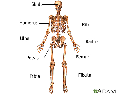
A pelvis x-ray is a picture of the bones in and around both hips. The pelvis connects the legs to the body.
x-ray
X-rays are a type of electromagnetic radiation, just like visible light. An x-ray machine sends individual x-ray waves through the body. The images...

How the Test is Performed
The test is done in a radiology department or in the health care provider's office by an x-ray technician.
You will lie down on the table. The pictures are then taken. You may have to move your body to other positions to provide different views.
How to Prepare for the Test
Tell the provider if you are pregnant. Remove all jewelry, especially around your belly and legs. You will wear a hospital gown.
How the Test will Feel
The x-rays are painless. Changing position may cause discomfort.
Why the Test is Performed
The x-ray is used to look for:
-
Fractures
Fractures
If more pressure is put on a bone than it can stand, it will split or break. A break of any size is called a fracture. If the broken bone punctures...
ImageRead Article Now Book Mark Article - Tumors
- Degenerative conditions of bones in the hips, pelvis, and upper legs
- Abnormal shape of your bones or joint
What Abnormal Results Mean
Abnormal results may suggest:
- Pelvic fractures
-
Arthritis of the hip joint
Arthritis
Arthritis is inflammation or degeneration of one or more joints. A joint is the area where 2 bones meet. There are more than 100 different types of...
 ImageRead Article Now Book Mark Article
 -
Tumors of the bones of the pelvis
Tumors
A bone tumor is an abnormal growth of cells within a bone. A bone tumor may be cancerous (malignant) or noncancerous (benign).
ImageRead Article Now Book Mark Article - Sacroiliitis (inflammation of the area where the sacrum joins the ilium bone)
-
Ankylosing spondylitis (abnormal stiffness of the spine and joint)
Ankylosing spondylitis
Ankylosing spondylitis (AS) is a chronic form of arthritis. It mostly affects the bones and joints at the base of the spine where it connects with t...
 ImageRead Article Now Book Mark Article
 - Arthritis of the lower spine
- Abnormality of the shape of your pelvis or hip joint
Risks
Children and the fetuses of pregnant women are more sensitive to the radiation of the x-ray. A protective shield may be worn over areas not being scanned or other imaging modalities can be used.
Reviewed By
Jason Levy, MD, FSIR, Northside Radiology Associates, Atlanta, GA. Also reviewed by David C. Dugdale, MD, Medical Director, Brenda Conaway, Editorial Director, and the A.D.A.M. Editorial team.
Beebe MJ. Fractures of the acetabulum and pelvis. In: Azar FM, Beaty JH, eds. Campbell's Operative Orthopaedics. 14th ed. Philadelphia, PA: Elsevier; 2021:chap 56.
Kapoor G, Toms AP. Current status of imaging of the musculoskeletal system. In: Adam A, Dixon AK, Gillard JH, Schaefer-Prokop CM, eds. Grainger & Allison's Diagnostic Radiology: A Textbook of Medical Imaging. 7th ed. Philadelphia, PA: Elsevier; 2021:chap 38.
Williams KD. Spondylolisthesis. In: Azar FM, Beaty JH, eds. Campbell's Operative Orthopaedics. 14th ed. Philadelphia, PA: Elsevier; 2021:chap 40.
Disclaimer
 All rights reserved.