X-ray - skeleton
A skeletal x-ray is an imaging test used to look at your bones. It is used to detect fractures, tumors, or conditions that cause wearing away (degeneration) of the bone.
x-ray
X-rays are a type of electromagnetic radiation, just like visible light. An x-ray machine sends individual x-ray waves through the body. The images...

Fractures
If more pressure is put on a bone than it can stand, it will split or break. A break of any size is called a fracture. If the broken bone punctures...
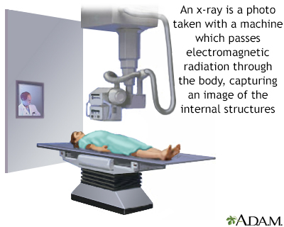
How the Test is Performed
The test is done in a hospital radiology department or in your health care provider's office by an x-ray technologist.
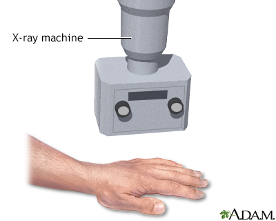
You will lie on a table or stand in front of the x-ray machine, depending on the bone that is injured. You may be asked to change position so that different x-ray views can be taken.
The x-rays pass through your body. A computer or special film records the images.
Structures that are dense (such as bone) will block most of the x-ray particles. These areas will appear white. Metal and contrast media (special dye used to highlight areas of the body) will also appear white. Structures containing air will be black. Muscle, fat, and fluid will appear as shades of gray.
How to Prepare for the Test
Tell your provider if you are pregnant. You must remove all jewelry before the x-ray.
How the Test will Feel
The x-rays are painless. Changing positions and moving the injured area for different x-ray views may be uncomfortable. If the whole skeleton is being imaged, the test most often takes 1 hour or more.
Why the Test is Performed
This test is used to look for:
- Fractures or broken bone
- Cancer that has spread to other areas of the body
-
Osteomyelitis (inflammation of the bone caused by an infection)
Osteomyelitis
Osteomyelitis is a bone infection. It is caused by bacteria or other germs.
ImageRead Article Now Book Mark Article - Bone damage due to trauma (such as an auto accident) or degenerative conditions
- Abnormalities in the soft tissue around the bone
Video Transcript
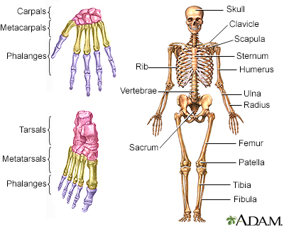
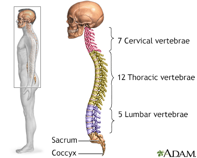
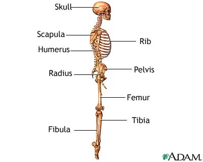
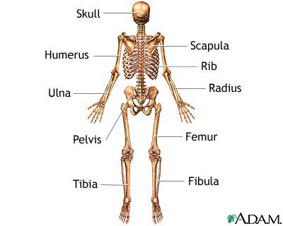
Skeletal system components - Animation
The skeletal system consists of approximately 206 bones, providing the body with structure and support. Let’s take a tour of various components that form the skeletal system. Here’s the skull. It has 8 cranial bones that protect the brain. The facial skeleton has 14 bones that provide a framework for the eye sockets, jaws, and teeth. The facial bones provide the framework for the various structures of the face including the overlying muscles, fat and skin. The vertebral column is composed of 24 individual vertebrae, along with two sets of fused bones called the sacrum and coccyx. In addition to providing support for the trunk of the body, the vertebral column protects the spinal cord. All together, there are 7 cervical, or neck vertebrae; 12 thoracic, or upper back, vertebrae; and 5 lumbar, or lower back, vertebrae. The sacrum is composed of 5 fused bones, while the coccyx, or tailbone, is typically made up of 3 to 5 bones. 12 pairs of ribs form a protective cage for the heart, lungs, and other internal organs. The first 7 ribs are called true ribs because they attach to the breastbone, or sternum. Ribs 8 through 12 are called false ribs, because they either attach indirectly, or, as in the case with ribs 11 and 12, float and don’t attach to the sternum at all. Now let’s take a look at the pair of shoulder blades, or scapulae, and the collar bones, or clavicles. It is very important for the scapulae to be mobile, because they connect to the shoulder joint, which is the most movable joint in the body. The bones of the upper limb include the humerus, which connects the shoulder with the elbow, the ulna, the radius, the wrist bones or carpals, the hand bones or metacarpals, and the finger bones or phalanges. To complete our tour, let’s take a look at the pelvic girdle, knee, and foot. The pelvic girdle is formed by a pair of hip bones. Each hip bone is comprised of 3 fused bones, the ilium, ischium, and pubis. The pelvic girdle connects with the femur or thigh bone at the hip joint. The femur is the longest bone in the body and is important for bearing the body’s weight while standing. At the knee, the femur articulates with the tibia or shin bone. The fibula does not bear weight, but several muscles attach to it. The patella, or kneecap, is suspended within muscle tendons and glides over the femur and tibia when the knee bends. And last, but certainly not the least, are the feet. The foot bones, which include the tarsals, metatarsals, and phalanges, are organized into a series of arches that allow the feet to support the body’s weight.
What Abnormal Results Mean
Abnormal findings include:
- Fractures
-
Bone tumors
Bone tumors
A bone tumor is an abnormal growth of cells within a bone. A bone tumor may be cancerous (malignant) or noncancerous (benign).
ImageRead Article Now Book Mark Article - Degenerative bone conditions
- Osteomyelitis
Risks
There is low radiation exposure. X-rays machines are set to provide the smallest amount of radiation exposure needed to produce the image. Most experts feel that the risk is low compared with the benefits.
Children and the fetuses of pregnant women are more sensitive to the risks of the x-ray. A protective shield may be worn over areas not being scanned.
Reviewed By
Linda J. Vorvick, MD, Clinical Professor Emeritus, Department of Family Medicine, UW Medicine, School of Medicine, University of Washington, Seattle, WA. Also reviewed by David C. Dugdale, MD, Medical Director, Brenda Conaway, Editorial Director, and the A.D.A.M. Editorial team.
Contreras F, Perez J, Jose J. Imaging overview. In: Miller MD, Thompson SR, eds. DeLee, Drez, & Miller's Orthopaedic Sports Medicine. 5th ed. Philadelphia, PA: Elsevier; 2020:chap 7.
Kapoor G, Toms AP. Current status of imaging of the musculoskeletal system. In: Adam A, Dixon AK, Gillard JH, Schaefer-Prokop CM, eds. Grainger & Allison's Diagnostic Radiology. 7th ed. Philadelphia, PA: Elsevier; 2021:chap 38.
Disclaimer

 All rights reserved.