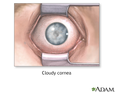
Cloudy cornea
A cloudy cornea is a loss of transparency of the cornea.
Causes
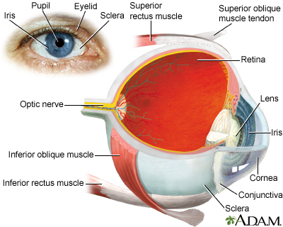
The cornea is the front wall of the eye. It is normally clear. It helps focus the light entering the eye.
Causes of cloudy cornea include:
- Inflammation of the cornea
- Sensitivity to non-infectious bacteria or toxins
- Infection of the cornea
- Keratitis
- Trachoma
- River blindness
-
Corneal ulcers
Corneal ulcers
The cornea is the clear tissue at the front of the eye. A corneal ulcer is an open sore in the outer layer of the cornea. It is often caused by inf...
 ImageRead Article Now Book Mark Article
 - Swelling (edema)
- Acute glaucoma
Glaucoma
Glaucoma is a group of eye conditions that can damage the optic nerve. This nerve sends the images you see to your brain. Most often, optic nerve da...
 ImageRead Article Now Book Mark Article
 - Birth injury
- Fuchs dystrophy
- Dryness of the eye due to Sjögren syndrome, vitamin A deficiency, or LASIK eye surgery
Sjögren syndrome
Sjögren syndrome is an autoimmune disorder in which the glands that produce tears and saliva are destroyed. This causes dry mouth and dry eyes. The...
 ImageRead Article Now Book Mark Article
 - Corneal dystrophy (inherited metabolic disease)
- Keratoconus
- Injury to the eye, including chemical burns and welding injury
- Tumors or growths on the eye
- Pterygium
- Bowen disease
Clouding may affect all or part of the cornea. It leads to different amounts of vision loss. You may not have any symptoms in the early stages.
Vision loss
Blindness is a lack of vision. It may also refer to a loss of vision that cannot be corrected with glasses or contact lenses. Partial blindness mean...

Home Care
Contact your health care provider. There is no appropriate home care.
When to Contact a Medical Professional
Contact your provider if:
- The outer surface of your eye appears cloudy.
- You have trouble with your vision.
Note: You will need to see an ophthalmologist (eye doctor) for vision or eye problems. However, your primary care provider may also be involved if the problem could be due to a whole-body (systemic) disease.
What to Expect at Your Office Visit
The provider or eye doctor will examine your eyes and ask about your medical history. The two main questions will be if your vision is affected and if you have seen a spot on the front of your eye.
Other questions may include:
- When did you first notice this?
- Does it affect both eyes?
- Do you have trouble with your vision?
- Is it constant or intermittent?
- Do you wear contact lenses?
- Is there any history of injury to the eye?
- Has there been any discomfort? If so, is there anything that helps?
Tests may include:
- Biopsy of eyelid tissue
- Computer mapping of the cornea (corneal topography)
- Schirmer test for eye dryness
- Special photographs to measure the cells of the cornea
-
Standard eye exam
Standard eye exam
A standard eye exam is a series of tests done to check your vision and the health of your eyes. This exam is performed by an ophthalmologist or opto...
 ImageRead Article Now Book Mark Article
 -
Ultrasound to measure corneal thickness
Ultrasound
Ultrasound uses high-frequency sound waves to make images of organs and structures inside the body.
 ImageRead Article Now Book Mark Article
Reviewed By
Franklin W. Lusby, MD, Ophthalmologist, Lusby Vision Institute, La Jolla, CA. Also reviewed by David C. Dugdale, MD, Medical Director, Brenda Conaway, Editorial Director, and the A.D.A.M. Editorial team.
Cioffi GA, Liebmann JM. Diseases of the visual system. In: Goldman L, Cooney KA, eds. Goldman-Cecil Medicine. 27th ed. Philadelphia, PA: Elsevier; 2024:chap 391.
Kataguiri P, Kenyon KR. Corneal and external eye manifestations of systemic disease. In: Yanoff M, Duker JS, eds. Ophthalmology. 6th ed. Philadelphia, PA: Elsevier; 2023:chap 4.25.
Kuborn AM, Hassan SE. The impact of vision loss on attitudes toward autonomous vehicles: a vision-centric analysis. Optom Vis Sci. 2024 ;101(6):424-34. PMID: 38990241 pubmed.ncbi.nlm.nih.gov/38990241/.
Patel SS, Zaguia F, Goldstein DA. Episcleritis and scleritis. In: Yanoff M, Duker JS, eds. Ophthalmology. 6th ed. Philadelphia, PA: Elsevier; 2023:chap 4.11.
Wang EY, Kong X, Wolle M, et al. Global trends in blindness and vision impairment resulting from corneal opacity 1984–2020: A meta-analysis. Ophthalmology. 2023:130(8):863-71.PMID: 36963570 pubmed.ncbi.nlm.nih.gov/36963570/.
Disclaimer
 All rights reserved.