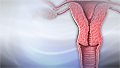
Cervix
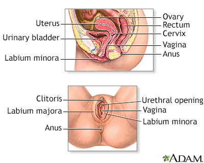
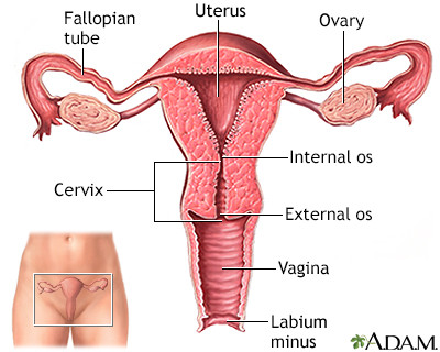
The cervix is the lower end of the womb (uterus). It is at the top of the vagina. It is about 2.5 to 3.5 centimeters (1 to 1.3 inches) long. The cervical canal passes through the cervix. It allows blood from a menstrual period and a baby (fetus) to pass from the womb into the vagina. Sperm travel from the vagina up the cervical canal into the uterine cavity, then into the fallopian tubes to fertilize the egg.
Vagina
The vagina is the female body part that connects the womb (uterus) and cervix to the outside of the body.

Conditions that affect the cervix include:
-
Cervical cancer
Cervical cancer
Cervical cancer is cancer that starts in the cervix. The cervix is the lower part of the uterus (womb) that opens at the top of the vagina.
 ImageRead Article Now Book Mark Article
 - Cervical infection
-
Cervical inflammation
Cervical inflammation
Cervicitis is swelling or inflamed tissue of the end of the uterus (cervix).
ImageRead Article Now Book Mark Article -
Cervical intraepithelial neoplasia (CIN) or dysplasia
Cervical intraepithelial neoplasia
Cervical dysplasia refers to abnormal changes in the cells on the surface of the cervix. The cervix is the lower part of the uterus (womb) that open...
ImageRead Article Now Book Mark Article -
Cervical polyps
Cervical polyps
Cervical polyps are fingerlike growths on the lower part of the uterus that connects with the vagina (cervix).
ImageRead Article Now Book Mark Article -
Cervical pregnancy
Cervical pregnancy
An ectopic pregnancy is a pregnancy that occurs outside the womb (uterus).
 ImageRead Article Now Book Mark Article
 - Cervical incompetence in pregnancy
Cervical cancer screening involves a Pap smear and an HPV test. For both these tests, the cells are taken from the cervix. A Pap test checks for premalignant (precancerous) changes in the cervix, and the HPV test checks for infection with human papillomavirus (HPV) that may lead to cervical cancer.
Pap smear
The Pap test mainly checks for changes in the cervix that may turn into cervical cancer. Cells scraped from the opening of the cervix are examined u...
HPV test
The HPV test is used to check for infection with HPV types associated with cervical cancer. Typically, the test looks for 14 different HPV types. H...
Reviewed By
Linda J. Vorvick, MD, Clinical Professor Emeritus, Department of Family Medicine, UW Medicine, School of Medicine, University of Washington, Seattle, WA. Also reviewed by David C. Dugdale, MD, Medical Director, Brenda Conaway, Editorial Director, and the A.D.A.M. Editorial team. Editorial update 08/12/2025.
Baggish MS. Anatomy of the cervix. In: Baggish MS, Karram MM, eds. Atlas of Pelvic Anatomy and Gynecologic Surgery. 5th ed. Philadelphia, PA: Elsevier; 2021:chap 42.
Cervix. Taber's Cyclopedic Medical Dictionary. 24th ed. F.A. Davis Company; 2021. www.tabers.com/tabersonline/view/Tabers-Dictionary/763678/all/cervix. Accessed December 17, 2024.
National Cancer Institute website. NCI dictionaries. Cervix. www.cancer.gov/publications/dictionaries/cancer-terms/def/cervix. Accessed January 7, 2025.
National Cancer Institute website. Cervical cancer screening. www.cancer.gov/types/cervical/screening. Updated February 13, 2025. Accessed August 12, 2025.
Disclaimer
 All rights reserved.