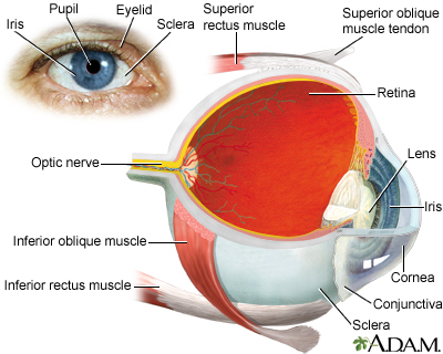
Browse A-Z

More <
bookmarks-menu

Sclera

The sclera is the white outer coating of the eye. It is tough, fibrous tissue that extends from the cornea (the clear front section of the eye) to the optic nerve at the back of the eye. The sclera gives the eyeball its white color. The cornea and sclera are made of the same type of collagen fibers. In the cornea, the fibers are arranged in sheets and layers which makes the cornea clear. In the sclera, the fibers are arranged randomly.

Review Date: 8/4/2023

Reviewed By

Franklin W. Lusby, MD, Ophthalmologist, Lusby Vision Institute, La Jolla, CA. Also reviewed by David C. Dugdale, MD, Medical Director, Brenda Conaway, Editorial Director, and the A.D.A.M. Editorial team.

References

Standring S. Eye. In: Standring S, ed. Gray's Anatomy: The Anatomical Basis of Clinical Practice. 42nd ed. Philadelphia, PA: Elsevier; 2021:chap 45.

Taber's Online. Sclera. www.tabers.com/tabersonline/view/Tabers-Dictionary/748617/all/sclera. Accessed September 29, 2023.

Disclaimer

The information provided herein should not be used during any medical emergency or for the diagnosis or treatment of any medical condition. A licensed medical professional should be consulted for diagnosis and treatment of any and all medical conditions. Links to other sites are provided for information only -- they do not constitute endorsements of those other sites. No warranty of any kind, either expressed or implied, is made as to the accuracy, reliability, timeliness, or correctness of any translations made by a third-party service of the information provided herein into any other language. © 1997- 2025 A.D.A.M., a business unit of Ebix, Inc. Any duplication or distribution of the information contained herein is strictly prohibited.
Images
Eye - Illustration Thumbnail

Eye

The eye is the organ of sight, a nearly spherical hollow globe filled with fluids (humors). The outer layer (sclera, or "white of the eye", and cornea) is fibrous and protective. The middle layer (choroid, ciliary body and the iris) is vascular. The innermost layer (retina) is sensory nerve tissue that is light sensitive. The fluids in the eye are divided by the lens into the vitreous humor (behind the lens) and the aqueous humor (in front of the lens). The lens itself is flexible and suspended by ligaments which allow it to change shape to focus light on the retina, which is composed of sensory neurons.

Illustration


URAC Seal
Health Content Provider
06/01/2025
A.D.A.M., Inc. is accredited by URAC, for Health Content Provider (www.urac.org). URAC's accreditation program is an independent audit to verify that A.D.A.M. follows rigorous standards of quality and accountability. A.D.A.M. is among the first to achieve this important distinction for online health information and services. Learn more about A.D.A.M.'s editorial policy, editorial process and privacy policy. A.D.A.M. is also a founding member of Hi-Ethics. This site complied with the HONcode standard for trustworthy health information from 1995 to 2022, after which HON (Health On the Net, a not-for-profit organization that promoted transparent and reliable health information online) was discontinued.
© 1997-2025ADAM Company Logo All rights reserved.